By Dr Chris Covelli
Overview
Regional anaesthesia is an important component of modern perioperative care, providing effective analgesia while reducing opioid use and improving recovery. The use of ultrasound guidance has significantly improved the safety and accuracy of nerve blocks by allowing clinicians to visualise anatomical structures in real time. However, identifying nerves on ultrasound can still be challenging due to complex anatomy, variable image quality, and operator experience.
I have had the pleasure to experience new groundbreaking AI technologies that are emerging in the area of regional ultrasound guided anaesthesia. AI-assisted ultrasound systems analyse images in real time and highlight key anatomical structures such as nerves and vessels. Early research suggests these tools may improve anatomical recognition, support trainee learning, and enhance scanning performance.

Healthcare in Europe – “A breakthrough in real-time ultrasound guidance for regional anesthesia.”https://healthcare-in-europe.com/en/news/a-breakthrough-in-real-time-ultrasound-guidance-for-regional-anesthesia.html
Why Nerve Localisation Is Difficult
Ultrasound-guided regional anaesthesia has dramatically improved the safety and accuracy of nerve blocks. However, identifying nerves and anatomical landmarks on ultrasound remains challenging, particularly for inexperienced practitioners.
Several factors contribute to this difficulty. First, ultrasound images often provide limited contrast between nerves and surrounding tissues, making structures difficult to distinguish. Additionally, technical factors such as poor needle visibility, deep anatomical targets, or patient characteristics such as obesity can further complicate image interpretation.
Ultrasound-guided blocks require clinicians to simultaneously interpret sonographic anatomy, manipulate the probe, and guide the needle toward the target structure. This complex visuospatial task demands significant experience and pattern recognition. As a result, the learning curve for regional anaesthesia can be steep, and incorrect interpretation of anatomy may lead to complications such as vascular puncture, nerve injury, or block failure.
What Is AI-Assisted Ultrasound?
AI-assisted ultrasound refers to the use of machine learning algorithms to analyse ultrasound images and identify anatomical structures in real time.
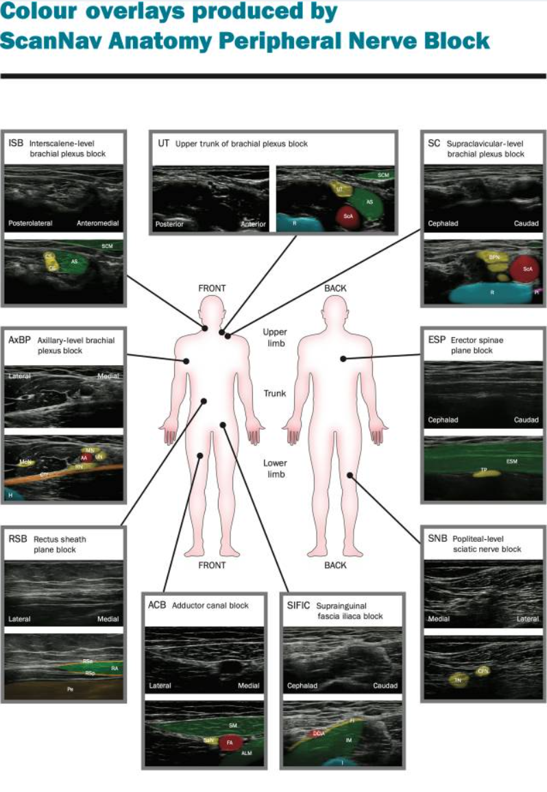
Most systems rely on deep learning neural networks trained on large datasets of annotated ultrasound images. These algorithms learn to recognise visual patterns associated with nerves, vessels, muscles, and fascial planes. Once trained, the system can analyse live ultrasound images and overlay colour highlights on key anatomical structures, helping clinicians interpret the image more easily.
In regional anaesthesia, AI tools may assist clinicians by:
-
Identifying nerves and surrounding anatomy
-
Highlighting critical safety structures such as vessels or pleura
-
Improving ultrasound image interpretation
-
Assisting with needle localisation and trajectory planning.
AI-assisted systems function as decision-support tools, augmenting the clinician’s interpretation rather than replacing clinical judgement. Their goal is to enhance anatomical recognition and improve procedural accuracy during nerve blocks.
Br J Anaesth. 2022 Aug 18;130(2):217–225. doi: 10.1016/j.bja.2022.06.031
What Does the Evidence Show?
Emerging research suggests that AI-assisted ultrasound may improve ultrasound scanning performance, particularly among less experienced clinicians.
A prospective study evaluating an assistive AI ultrasound device found that non-expert anaesthetists were more likely to obtain the correct block view when using AI assistance. In this study, the correct block view was obtained in 90.3% of scans with AI compared with 75.1% without AI assistance.
Similarly, correct identification of sonographic anatomical structures was significantly higher when AI support was used (88.8% vs 77.4% without AI).
These findings suggest that AI may enhance both image acquisition and interpretation, two critical steps in ultrasound-guided nerve blocks.
Broader reviews of the literature have also identified several potential benefits of AI-assisted ultrasound. These include improved identification of anatomical landmarks, enhanced visualisation of needle advancement, and optimisation of ultrasound image interpretation. AI tools may therefore help reduce complications such as injury to surrounding structures or incorrect needle placement.
However, despite promising early results, the current body of evidence remains relatively limited, and further large-scale clinical trials are required to confirm whether these technologies improve patient outcomes.
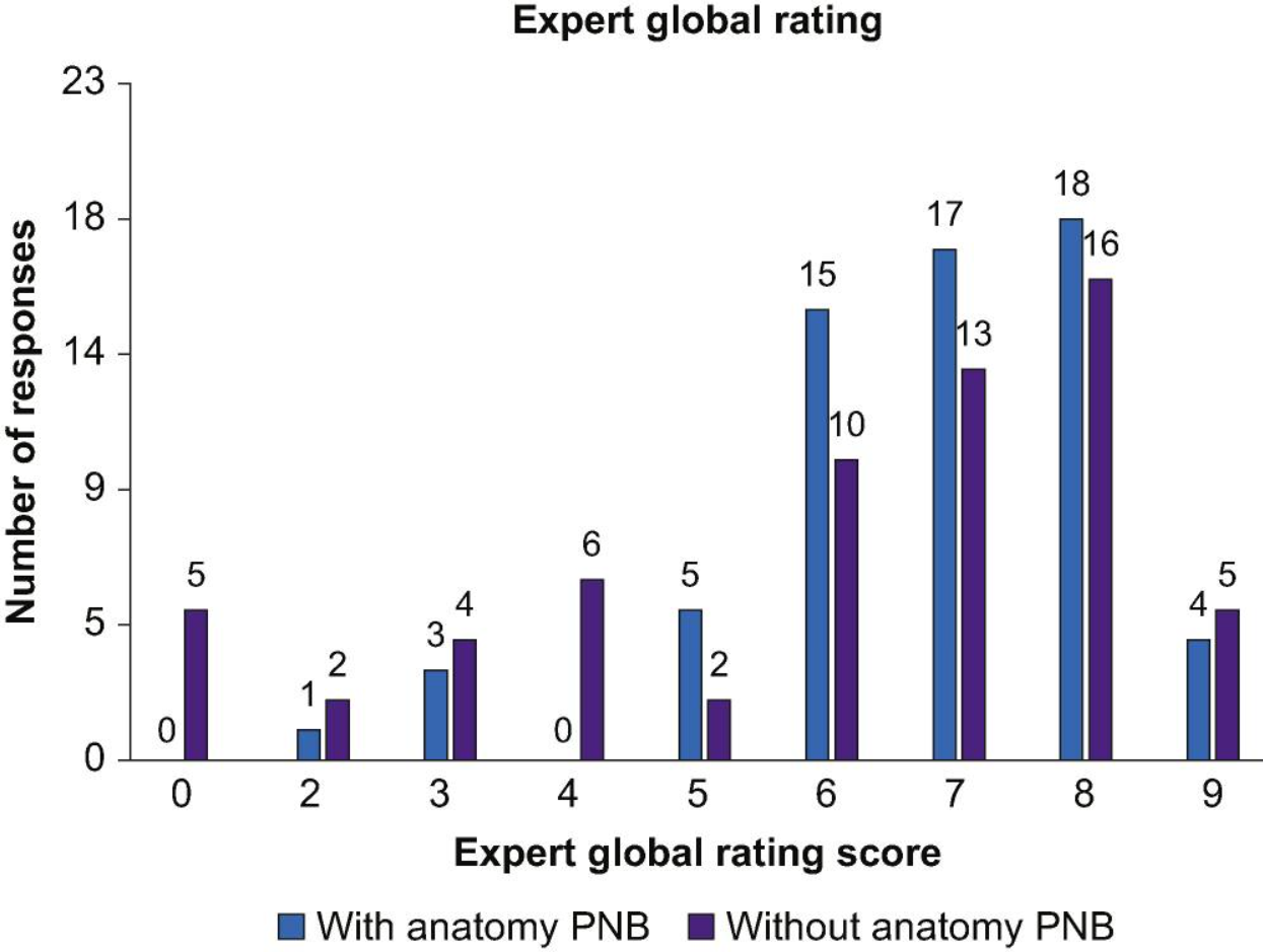
Expert global rating score. Distribution of all expert global rating scores, showing a breakdown of scans performed with or without ScanNav Anatomy Peripheral Nerve Block. PNB, Peripheral Nerve Block.
How AI Could Transform Regional Anaesthesia Training
One of the most exciting potential applications of AI-assisted ultrasound is in education and training.
Learning regional anaesthesia requires the development of strong sono-anatomy recognition skills, which traditionally develop through repeated scanning and expert supervision. AI tools could accelerate this learning process by providing real-time anatomical guidance during ultrasound scanning.
For trainees, AI systems may:
-
Help identify correct anatomical landmarks during scanning
-
Reinforce recognition of normal sonographic anatomy
-
Improve confidence during early learning stages
-
Provide immediate visual feedback.
Studies have suggested that AI assistance may help non-expert clinicians improve their ability to acquire correct ultrasound views and recognise anatomical structures, which could shorten the learning curve for ultrasound-guided blocks.
More broadly, AI-assisted ultrasound could become an important component of technology-enhanced regional anaesthesia training, alongside simulation, augmented reality, and ultrasound-guided training platforms.
Limitations and Concerns of AI-Assisted Ultrasound
Despite the enthusiasm surrounding AI-assisted ultrasound, several limitations and concerns remain.
First, the current evidence base is still developing. Reviews of the literature highlight that many studies involve small sample sizes or experimental settings, and high-quality randomised controlled trials are still lacking.
Another important concern is over-reliance on AI systems. While AI can assist with image interpretation, clinicians must still possess a strong understanding of ultrasound anatomy. Incorrect AI identification of anatomical structures could potentially mislead inexperienced users if they rely too heavily on the technology.
Technical limitations also remain. AI systems may struggle in cases with unusual anatomy, poor image quality, or deep anatomical structures, and their performance may vary depending on the ultrasound machine or scanning conditions.
Finally, there are broader issues related to regulation and standardisation of AI devices. Different AI systems may be evaluated using different datasets and performance metrics, making it difficult for clinicians to compare their accuracy and clinical utility.
Addressing these challenges will require collaboration between clinicians, engineers, and regulatory bodies to ensure that AI technologies are implemented safely and effectively in clinical practice.
Conclusion
Artificial intelligence is beginning to reshape how clinicians interpret ultrasound during regional anaesthesia. Early studies suggest that AI-assisted systems can improve the identification of anatomical structures and help clinicians obtain optimal ultrasound views, particularly among less experienced users. By highlighting nerves, vessels, and surrounding anatomy in real time, these technologies have the potential to enhance procedural accuracy and support the learning process for trainees.
However, AI should be viewed as a clinical decision-support tool rather than a replacement for anatomical knowledge or ultrasound expertise. The current evidence base remains limited, and further research is needed to determine whether these systems translate into improved patient outcomes and reduced complications. As the technology continues to evolve, AI-assisted ultrasound may become an increasingly valuable adjunct in regional anaesthesia practice and education.
References
Bowness JS, Burckett-St Laurent D, Hernandez N, Keane PA, Lobo C, Margetts S, et al. Assistive artificial intelligence for ultrasound image interpretation in regional anaesthesia: an external validation study. Br J Anaesth. 2023;130(2):217-225. doi:10.1016/j.bja.2022.06.031.
Bowness JS, Burckett-St Laurent D, Margetts S, Pawa A, Noble JA, Higham H, et al. Evaluation of the impact of assistive artificial intelligence on ultrasound scanning for regional anaesthesia. Br J Anaesth. 2023;130(2):226-233.
Bowness JS, Lobo C, Burckett-St Laurent D, Noble JA, Pawa A, Higham H, et al. Variability between human experts and artificial intelligence in identification of anatomical structures by ultrasound in regional anaesthesia: a framework for evaluation of assistive artificial intelligence. Br J Anaesth. 2024;132(5):1063-1072.
Healthcare in Europe. A breakthrough in real-time ultrasound guidance for regional anesthesia [Internet]. Healthcare in Europe; [cited 2026 Mar 17]. Available from: A breakthrough in real-time ultrasound guidance for regional anesthesia