By Zheng Cheng Zhu
Key reference:
Karamchandani et al. (2024). Tracheal intubation in critically ill adults with a physiologically difficult airway. An international Delphi study. Intensive care medicine, 50(10), 1563–1579. https://doi.org/10.1007/s00134-024-07578-2
Quick Summary
-
The “physiologically difficult airway” (PDA) was first coined by Mosier et al. in 2015, filling a conceptual gap in the management of airways in critically unwell patients.
-
Critically unwell patients experience higher rates of peri-induction adverse events (one in five patients) irrespective of first pass intubation success, underscoring PDA as an additional entity to the traditional “anatomical” difficult airway.
-
Hypoxaemia, hypotension, increased intracranial pressure, right ventricular failure, obesity and pregnancy present the most common and challenging PDA.
-
Complications mostly occur during induction, intubation and transition to positive pressure ventilation.
-
Environmental factors (ED, ICU, under-resourced ward) and human factors increase the risk of complications for PDAs
-
Recent consensus statements published by Society of Critical Care Anaesthesiologists provide guidance on the management of PDAs
Preamble
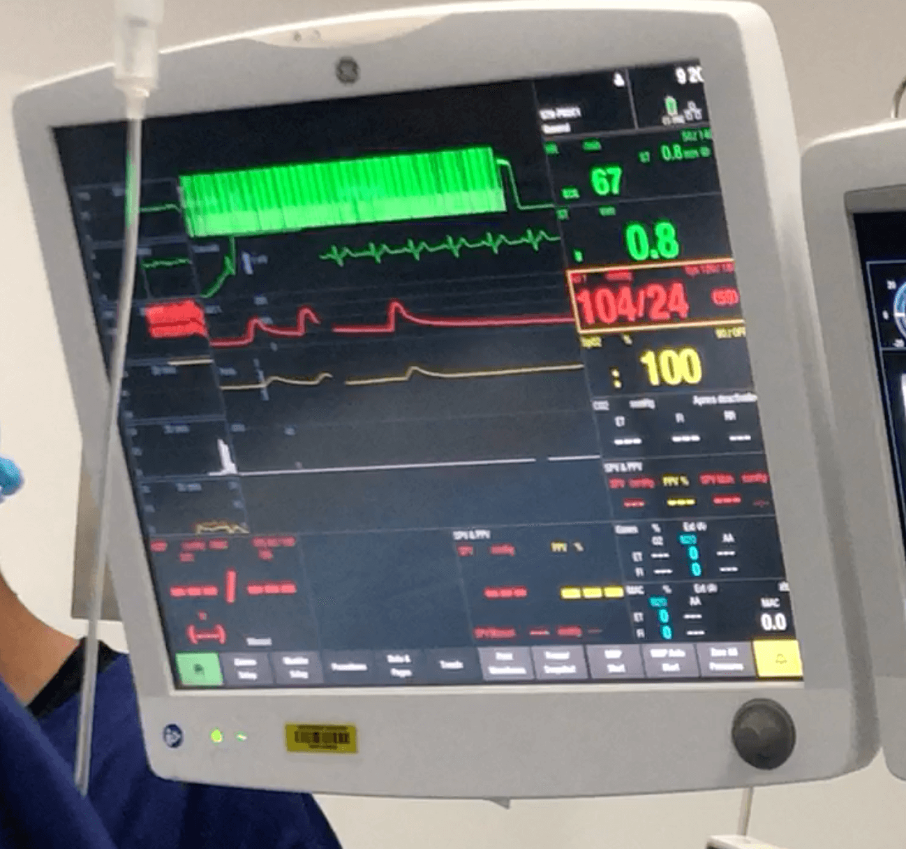
You are an overnight anaesthesia registrar. You receive a call from a panicked ICU registrar requesting your assistance with an emergency intubation. The patient is a 60-year-old male admitted to ICU for severe community-acquired pneumonia, who has now deteriorated with respiratory fatigue, respiratory acidosis, and desaturation to 90%, despite an FiO₂ of 60% on non-invasive ventilation. Invasive blood pressure monitoring shows a mean arterial pressure of 67 mmHg and a sinus tachycardia of 110 bpm, supported by 2 mcg/min of noradrenaline. You attend to the patient immediately.
You confirm the patient’s previous airway grade as a Grade I with direct laryngoscopy. On examination, he has a thyromental distance of 7 cm, satisfactory mouth opening, and good neck extension. He is adequately fasted.
“Should be an easy airway, right?”

Tracheal intubation remains one of the most specialised and high-risk procedures performed in critical care, requiring the clinician to navigate the technical challenges of airway manipulation, rapid physiological alterations associated with apnoea, induction agents, and initiation of positive pressure ventilation—all within a matter of seconds.
Traditionally, an airway is anticipated to be “difficult” when there are anatomical features that impede the ability to ventilate and oxygenate the patient using bag-mask ventilation, tracheal intubation, or rescue supraglottic airway devices. These limitations prolong apnoea time and increase the risk of peri-intubation complications, including the “can’t intubate, can’t oxygenate” (CICO) scenario. The “anatomically difficult airway” is a well-recognised entity that has driven extensive advances in airway equipment, practice guidelines, and training for its management.
However, improvements in first-pass success rates have uncovered persistent peri-intubation complication rates in the critically ill. Indeed, observational studies have consistently demonstrated serious complication rates—such as hypoxaemia, haemodynamic instability, and cardiac arrest—to be around 20% in critically ill patients undergoing intubation, despite first-pass success. The International Observational Study to Understand the Impact and Best Practices of Airway Management in Critically Ill Patients (INTUBE) found that 45.2% of the 2,964 ICU patients undergoing intubation experienced at least one major adverse peri-intubation event. Specifically, while first-pass success was imperative in avoiding further critical desaturation (p<0.001 for both two attempts and >two attempts), it was not shown to be protective against haemodynamic instability (p=0.416 for first pass vs two attempts, and p=0.572 for >two attempts). As such, pathophysiological alterations not only pose peri-intubation challenges independent of traditional anatomical limitations, but they also exacerbate the negative consequences of a failed first-pass attempt in patients with compromised physiological reserve.

Mosier and colleagues first coined the term “physiologically difficult airway” (PDA) in 2015, recognising that pre-existing physiological derangements increase the rates of serious complications during intubation and the transition to positive pressure ventilation. The concept emphasises the need for critical care specialists to consider specific physiological derangements—beyond airway anatomy—when planning an intubation. Since then, growing recognition and research have improved the understanding and management of the PDA. In 2024, an international Delphi study chaired by the Society of Critical Care Anesthesiologists (SOCCA) Physiologically Difficult Airway Task Force synthesised the best available evidence and expert consensus into practice statements to guide the safe intubation of PDAs.
This article will explore the concept of the PDA, specific physiological derangements we commonly encounter, and key recommendations from the SOCCA Delphi study to aid our management of the critically ill airway.
What is a “Physiologically Difficult Airway (PDA)”
Mosier’s group and SOCCA define PDA as the patient who presents with pre-intubation physiological or pathophysiological factors that increase the risk of peri-intubation adverse events despite one or few intubation attempts, irrespective of (and possibly exaggerate) the effect of an anatomically difficult airway.
Multiple large observational studies have demonstrated pre-intubation haemodynamic compromise, defined as hypotension with mean arterial pressure (MAP) <65mmHg, systolic blood pressure <130mmHg, or sepsis, and requirement for pharmacological augmentation (e.g. need for vasopressor or fluid bolus, diuresis, or avoidance of propofol) to be associated with post-intubation hypotension. Similarly, pre-intubation respiratory failure requiring non-invasive ventilation, emergency or cardiac indication for intubation, and fluid resuscitation are all associated with post-intubation hypoxaemia. Unsurprisingly, age and advanced disease grades are patient factors that increase the risk of both hypotension and hypoxaemia.
Induction, apnoea, intubation and positive pressure ventilation impose drastic physiological changes and demands upon patients’ compensatory reserve, which is evidently diminished and largely exhausted in the critically ill. Hypotension and hypoxaemia commonly ensue due to inability to overcome the haemodynamic effects of anaesthetic agents, impaired oxygenation, hypermetabolic state, and unfavourable cardiopulmonary biomechanics of positive pressure ventilation. An understanding of specific physiological derangements and their effects on peri-intubation physiology is therefore paramount to mitigating these risks.
Understanding specific PDA scenarios
In their first description of PDA in 2015, Mosier and colleagues described 4 commonly encountered PDAs. This has since expanded to 6 in the SOCCA Delphi study. We summarise their pathophysiology, optimisation strategies and their associated evidence below:
Hypoxaemia
Failure to maintain adequate arterial oxygenation and resultant tissue hypoxia is one of the most common indications for intubation, and one that paradoxically carries the most significant risk of peri-intubation desaturation and worsening hypoxia, haemodynamic instability, hypoxic brain injury, and arrest.
Type 1 respiratory failure (hypoxaemic respiratory failure) results from gross ventilation-perfusion (V/Q) mismatch from pulmonary shunting (V/Q <1) where blood passes through alveolar units without adequately participating in gaseous exchange. Common causes include pneumonia, pulmonary oedema and acute respiratory distress syndrome. When intubating, type 1 respiratory failure patients are more prone to desaturation due to impaired gaseous exchange and limited response to pre-oxygenation. For the critically ill, an increased O2 demand may rapidly deplete an already compromised O2 reserve, while patients in physiological extremes such as severe obesity, pregnancy, paediatric and the elderly have diminished functional residual capacity (FRC) to maintain a sufficient O2 reservoir. However, severe Type 2 respiratory failure, particularly from obstructive airway disease, can present far greater challenges in ventilation and oxygenation. Managing these patients requires a delicate balance to mitigate the risks of under-ventilation, barotrauma and dynamic hyperinflation.

Optimisation strategies
The goal for these patients is to ensure optimal pre-oxygenation, appropriate airway assessment, intubation and ventilator setup, to prolong safe apnoea time (time between apnoea to critical desaturation), minimise total apnoea time, and maximise first-pass success rate.
Preoxygenation aims to denitrogenise a patient’s FRC with O2 and create an O2 reserve during apnoea. Standard method of pre-oxygenation using non-rebreather mask or bag-valve mask at maximal O2 flow is often inadequate for the critically ill, as their elevated respiratory rate limits tidal volume, impairs lung recruitment, and increases entrained ambient air which dilutes FiO2. Non-invasive ventilation (NIV) partly circumvents the problem by providing a tighter seal, positive end-expiratory pressure (PEEP) to improve lung recruitment, reduce shunting and improve oxygenation, and pressure support for additional tidal volumes. A randomised controlled trial by Baillard et al. showed patients undergoing intubation with hypoxaemic respiratory failure experienced less adverse events and desaturation <80% (17.8% vs. 41.3%) when pre-oxygenated with NIV compared to bag-valve mask. Pre-intubation anxiolytics and sedation may be required for patients with altered conscious states to improve compliance with NIV.
Once in the apnoeic phase post-induction, one may further prolong safe apnoea time with apnoeic oxygenation or positive mask ventilation. Apnoeic oxygenation involves the continued delivery of low-flow or high flow O2 during apnoea, entraining O2 down the lungs via the diffusion gradient generated by the patient’s alveolar O2 uptake. There is mixed evidence of its efficacy prolonging safe apnoea time and prevention of severe hypoxaemia.
While positive pressure mask ventilation post-induction has been traditionally discouraged (contraindicated in “classic” rapid sequence induction) due to concerns of gastric insufflation and aspiration, it has seen increased adoption by critical care specialists for safely managing critically ill, hypoxic patients with compromised FRC and O2 reserve. Real-time sonographic studies have demonstrated that mask ventilation pressures of less than 15cmH2O can safely improve ventilation without causing significant gastric insufflation. Recent randomised controlled trial investigating 400 intubations in US ICUs showed that mask ventilation not only reduced rates of severe hypoxaemia by 52%, aspiration events were not increased compared to the no-ventilation group. Both strategies can be considered to delay desaturation.

Ultimately, all steps should be taken to maximise first pass success as to minimise total apnoea time. Unsurprisingly, first pass failure requiring multiple attempts is directly related to increased rates of severe hypoxia and haemodynamic instability. Optimal patient positioning with 30 degrees head ramping, careful airway assessment, use of video laryngoscopy, and in cases of predicted “anatomical difficult airway”, use of intubation adjuncts (bougie/stylet), hyperangulated blade, and BURP, are all protocolised practices that are familiar for critical care physicians.
Hypotension
The critically ill patients often display hypotension as a late-stage sign of haemodynamic decompensation and shock. All four types of shock (distributive, hypovolaemic, obstructive, cardiogenic) are worsened to some degree by the venodilation, vasodilation and negative inotropic effects of anaesthetic agents and haemodynamic alterations from the transition to positive pressure ventilation. Unsurprisingly, post-intubation haemodynamic instability is by far the most common adverse event as illustrated by the INTUBE study.
Optimisation strategies
Appropriate pre-induction resuscitation, haemodynamic support with vasopressors, and use of “cardiostable” induction can assist to minimise/prevent peri-intubation cardiovascular collapse.
Volume resuscitation is a common first-line strategy for correcting hypotension, although careful assessment of patient fluid balance and volume tolerance is needed to avoid iatrogenic overload and cardiopulmonary decompensation. The multicentre randomised PrePARE and subsequent PrePARE II trial analysed the effectiveness of initiating a 500ml crystalloid bolus prior to induction for the critically ill and found no effect in reducing rates of cardiovascular collapse. As such, indiscriminate administration of fluid bolus is discouraged. Rather, individualised selection should be made through assessing fluid responsiveness (straight leg raise, pulse pressure variation (PPV), echocardiogram-evident inferior vena cava (IVC) collapsibility and left ventricular dynamic volume assessments) and balancing against risk of overload (through clinical and echocardiogram assessment, chest x-ray etc.)
Vasopressor support may be used to dampen the effects of induction agents and positive pressure ventilation to maintain perfusion pressure. Evidence is mixed around use of prophylactic vasopressor to prevent peri-induction cardiovascular collapse. Nevertheless, patients with distributive and cardiogenic shock are likely to benefit from the reduction in post-induction vasoplegia.
Appropriate choices of induction agents and their dosages are paramount to minimise haemodynamic instability during induction. Propofola nd thiopentone causes loss of vascular tone and bradycardia, leading to reduced preload, afterload and coronary perfusion pressure. These agents are therefore avoided in critically ill patients in favour of more “cardiac stable” agents such as etomidate, midazolam, ketamine, and fentanyl co-induction. There is ongoing debate regarding the superiority of either etomidate or ketamine in the critically ill in reducing cardiovascular instability. Regardless, one should judiciously dose-reduce based on patient factors and severity of illness, or employ an induction strategy to limit doses required of each agent to achieve sufficient anaesthesia.

Right heart failure
In addition to haemodynamic instability, Mosier specifically distinguished moderate-severe right ventricular (RV) dysfunction and failure as a PDA entity. Rightfully so, RV physiology is intimately related and exquisitely sensitive to the physiological alterations caused by anaesthetic induction and positive pressure ventilation.
In normal right heart circulation, the RV is a low pressure, high compliance, flow-based chamber that mobilises venous blood through the pulmonary vasculature. It is preload dependent, reliant on adequate venous return to generate Frank-Starling mediated contractility, and afterload sensitive, where marginal increases in pulmonary pressure in a low-pressure system can cause RV strain and trigger RV compensation. Long-standing pulmonary hypertension (from pulmonary vascular disease, left ventricular dysfunction, chronic airway disease, or chronic pulmonary embolism) leads to pathological RV remodelling, RV dysfunction, and ultimately RV failure, where the RV can no longer overcome excess afterload, resulting in retrograde flow, reduced left heart and coronary perfusion, and cardiovascular collapse.
Unfortunately, induction and positive pressure ventilation can impair RV compensatory mechanisms and add further RV strain
-
Vasodilatory effects of induction agents reduce venous pressure and preload;
-
Transient hypoxaemia and hypercapnia during apnoea results in pulmonary vasoconstriction and increase in pulmonary vascular resistance;
-
Positive pressure ventilation increases intrathoracic pressure with a net effect of impeding venous return and increasing pulmonary vascular resistance, causing both reduced preload and increased RV afterload.
As such, intubating a patient with RV failure can result in RV decompensation and cardiovascular collapse.
Optimisation strategies
Optimisation of these patients requires a multidisciplinary approach and careful assessment. Common ventilatory aims would be to ensure adequate pre-oxygenation with sufficient etO2 using low PEEP NIV strategy, shorten apnoea time with apnoeic oxygenation to avoid hypoxaemia, and ventilate using spontaneous breathing modes to minimise positive pressure. Haemodynamics often needs to be evaluated using bedside echocardiograms to determine RV strain, contractile reserve and likelihood for fluid responsiveness. Use of fluid, vasopressors, and introduction of pulmonary vasodilators such as inhaled nitrous oxide should be guided by an expert team.
Intracranial hypertension
Intracranial pressure (ICP) is modelled by the Monro-Kellie doctrine. During cerebrovascular insults, such as intracranial haemorrhage, malignant ischaemic stroke, traumatic brain injury, and meningoencephalitis, intracranial pressure rises due to excess volume of blood and cerebral oedema within the rigid calvarium, resulting in compromised cerebral perfusion pressure and risk of herniation syndromes.
Laryngoscopy can cause a significant spike in ICP and worsen cerebral insult. Laryngoscopy may directly stimulate the gag/cough reflex in under-anaesthetised patients, in addition to the well-recognised sympathetic reflex that causes drastic spikes in heart rate, blood pressure and consequently raised ICP.

Optimisation strategies:
Key aims in the peri-intubation phase include pre-intubation optimisation, blunting of reflex sympathetic response to laryngoscopy, and post-intubation ventilation strategy.
In addition to standard preparation of pre-oxygenation and haemodynamic stabilisation, patient should be optimised to reduce ICP as much as possible
-
Ventilate to etCO2 target that correlate with PaCO2 of 35-40 to limit cerebral vasodilation;
-
Manage hypertension with short-acting beta-blockers and/or analgesia;
-
Non-pharmacological management (head-up 30 degrees, minimise neck compression, hyperthermia avoidance) and pharmacological (hypertonic saline if clinical evidence of worsening ICP);
-
Light sedation may be necessary to manage the agitated/non-compliant patient.
The neurocritical induction is heavily geared to blunting of the sympathetic reflex using large dose opioids. Fentanyl 3-5mcg/kg 1-3min prior to laryngoscopy is most commonly used in the emergent setting, while anaesthetists may have the luxury of accessing faster-onset and titratable opioids such as alfentanil and remifentanil.
Pharmacological strategies should not distract from non-pharmacological steps to minimise laryngoscopy manipulation:
-
Maximise first pass success as previously discussed;
-
Minimise force used with VL to achieve glottic exposure.
Once transitioned to positive pressure ventilation, neuroprotective ventilation strategy should be adopted:
-
Avoid hypoxaemia
-
Ventilate to PaCO2 30-35mmHg;
-
Minimal PEEP to avoid ICP increases;
-
Paralysis may be considered to further reduce ICP
Obesity & Pregnancy
Obesity and pregnancy have been specifically added by the SOCCA Delphi study as two PDAs. The predominant concern surrounds the restricted FRC, increased V/Q mismatch, and reduced O2 reserve due to increased metabolic demand, which limit the effectiveness of pre-oxygenation and make positive pressure ventilation particularly challenging. Haemodynamic effects of induction may be poorly tolerated from diminished compensatory reserve, or underlying cardiomyopathy. Lastly, the obese and parturient patients are at increased risk of aspiration during induction.
Optimisation strategies
While these patients present significant challenges in the peri-intubation setting, a structured approach using strategies outlined in “hypoxaemia” and “hypotension” can be similarly used to limit adverse events. Specifically, adequate pre-oxygenation, optimal patient positioning with ramping and a right wedge to prevent aortocaval compression, and careful haemodynamic monitoring with adequate resuscitation are important considerations.

Human factors in managing PDA
PDA adds additional cognitive and logistical challenges to an already complex procedure. Practitioners are often faced with high-acuity, time-sensitive and hyperdynamic scenarios in managing rapidly deteriorating patients, which can be overwhelming even for the most seasoned, leading to mistakes. Crisis resource management (CRM), which are non-technical skills that allow for optimal organisation and utilisation of available skillset, manpower and equipment in crisis situations, is paramount to enhance teamwork and performance.
Some key principles of CRM include clear role delegation appropriate for level of experience and training, closed loop communication, effective team leadership and shared mental model, all of which have been incorporated in airway management guidelines and simulation training.
The use of an “airway checklist” is common practice in ED and ICUs, providing a cognitive aid to ensure comprehensive patient, personnel, equipment and environmental preparation and minimise critical omissions prior to embarking on induction.
The team should at a minimum compose of a team leader who has overview of patient progress and provide clear decision-making, a primary airway operator, and an airway assistant/second airway operator. Clear verbalisation of airway and medication plan, including explicit checkpoints to enact contingency plans and seek early help, helps to establish a shared mental model of priorities and goals, catch complications early, and prevent task fixation and mobilise help to minimise further deteriorations.
Key SOCCA Statements
The following table summarises the key statements from the Delphi study on the approach to managing a PDA:
-
Team preparation
-
Use of airway checklists,
-
Team assignment with at least 3 healthcare workers,
-
Crisis resource management, including clear team roles, communication loops, shared mental model, cognitive aids etc.,
-
Simulation-based training.
Patient preparation and optimisation
-
Routinely perform airway assessment to anticipate difficult anatomical airway,
-
Haemodynamic stabilisation with interventions such as vasopressor or inotrope infusion,
-
Use of point-of-care ultrasound to assess and appropriately manage cardiac-related compromise,
-
Use of non-invasive ventilation pre-oxygenation, apnoeic oxygenation, gentle positive pressure mask ventilation to avoid desaturation.
Performing the rapid sequence induction (RSI)
-
Use of the “head up” 30 degrees laryngoscopy position
-
Use of a “modified” RSI with dose-adjusted rapid onset hypnotic (propofol, ketamine or etomidate), rapid-acting neuromuscular blocker (suxamethonium or rocuronium), judicious use of positive-pressure mask ventilation to optimise intubating conditions,
-
Use of video laryngoscopy with bougie/stylet should be used routinely during the first attempt.
Post-intubation care
-
Immediate priorities include confirmation of tube placement (consistent etCO2 pattern over 7 breaths) and management of complications, most commonly cardiovascular instability and hypoxaemia,
-
Use of lung protective ventilation, using
-
Tidal volumes 6-8ml/kg of predicted body weight (PBW)
-
PEEP >= 5 cmH2O
-
Plateau pressure <30 cmH2O
-
FiO2 titrated to SpO2 aims 92-95%
-
-
Invasive blood pressure monitoring, central venous access to manage persistent haemodynamic instability post-intubation.
-
Conclusion
Despite the patient having no red flags for difficult laryngoscopy, you recognise the patient has a physiologically difficult airway. Together with your ICU colleagues, you set out to maximise preparations to optimise your attempt
-
You organised your team with the ICU reg as the team leader, yourself as the primary airway operator, and the nurse-in-charge as the airway assistant, with the resident performing drug administration.
-
Your team contacts your respective consultant-on-call to be readily available to assist if the patient deteriorates.
-
You verbalise your plan to the team, with plan A being ETT size 8 with size 4 VL with bougie +/- BURP (max 2 attempts), plan B for second generation size 4 LMA + consultant assistance, with plan C to revert to BMV if plan B fails or saturation <88% at any stage and consider plan D – front of neck access.
-
During this time, you positioned the patient to head up 30 degrees, pre-oxygenate the patient with NIV on 100% FiO2 for 5 min and provided a 500ml crystalloid bolus which increased the patient’s MAP to 72mmHg.
-
You opted to give a cardiac-stable modified RSI of 1mg/kg ketamine, 2mg midazolam and 1.2mg/kg rocuronium, with gentle positive mask ventilation post induction.
Thankfully, your first pass was successful. The patient’s saturation decreased to 95%, blood pressure was relatively stable on 5mcg/min norad.
While advances in training, guidelines and airway technologies have increased our competencies in managing the anatomically difficult airways, the PDA has only gained recognition over the last decade, unveiling the multidimensional complexities in its management. This recent Dephi study by SOCCA provides guidance on best practice of managing PDAs, and provides a robust foundation for ongoing research regarding their feasibility in clinical practice.
References
Al-Saadi, M. A., Heidari, B., Donahue, K. R., Shipman, E. M., Kinariwala, K. N., & Masud, F. N. (2023). Pre-Existing Right Ventricular Dysfunction as an Independent Risk Factor for Post Intubation Cardiac Arrest and Hemodynamic Instability in Critically Ill Patients: A Retrospective Observational Study. Journal of intensive care medicine, 38(2), 169–178. https://doi.org/10.1177/08850666221111776
Baillard, C., Prat, G., Jung, B., Futier, E., Lefrant, J. Y., Vincent, F., Hamdi, A., Vicaut, E., & Jaber, S. (2018). Effect of preoxygenation using non-invasive ventilation before intubation on subsequent organ failures in hypoxaemic patients: a randomised clinical trial. British journal of anaesthesia, 120(2), 361–367. https://doi.org/10.1016/j.bja.2017.11.067
Bouvet, L., Albert, M. L., Augris, C., Boselli, E., Ecochard, R., Rabilloud, M., Chassard, D., & Allaouchiche, B. (2014). Real-time detection of gastric insufflation related to facemask pressure-controlled ventilation using ultrasonography of the antrum and epigastric auscultation in nonparalyzed patients: a prospective, randomized, double-blind study. Anesthesiology, 120(2), 326–334. https://doi.org/10.1097/ALN.0000000000000094
Casey, J. D., Janz, D. R., Russell, D. W., Vonderhaar, D. J., Joffe, A. M., Dischert, K. M., Brown, R. M., Zouk, A. N., Gulati, S., Heideman, B. E., Lester, M. G., Toporek, A. H., Bentov, I., Self, W. H., Rice, T. W., Semler, M. W., & PreVent Investigators and the Pragmatic Critical Care Research Group (2019). Bag-Mask Ventilation during Tracheal Intubation of Critically Ill Adults. The New England journal of medicine, 380(9), 811–821. https://doi.org/10.1056/NEJMoa1812405
Karamchandani, K., Nasa, P., Jarzebowski, M., Brewster, D. J., De Jong, A., Bauer, P. R., Berkow, L., Brown, C. A., 3rd, Cabrini, L., Casey, J., Cook, T., Divatia, J. V., Duggan, L. V., Ellard, L., Ergan, B., Jonsson Fagerlund, M., Gatward, J., Greif, R., Higgs, A., Jaber, S., … Society of Critical Care Anesthesiologists (SOCCA) Physiologically Difficult Airway Task Force (2024). Tracheal intubation in critically ill adults with a physiologically difficult airway. An international Delphi study. Intensive care medicine, 50(10), 1563–1579. https://doi.org/10.1007/s00134-024-07578-2
Mosier, J. M., Joshi, R., Hypes, C., Pacheco, G., Valenzuela, T., & Sakles, J. C. (2015). The Physiologically Difficult Airway. The western journal of emergency medicine, 16(7), 1109–1117. https://doi.org/10.5811/westjem.2015.8.27467
Mosier, J. (2024). The Physiologically Difficult Airway and Management Considerations. Curr Anesthesiol Rep 14, 446–457 . https://doi.org/10.1007/s40140-024-00629-w
Myatra, S. N., Divatia, J. V., & Brewster, D. J. (2022). The physiologically difficult airway: an emerging concept. Current opinion in anaesthesiology, 35(2), 115–121. https://doi.org/10.1097/ACO.0000000000001102
Nickson, C. (2023). Intubation of the Neurocritical Care Patient. Life In the Fast Lane.URL: https://litfl.com/intubation-of-the-neurocritical-care-patient/, [Last accessed 25/11/2024]
Patel, S. D., & Habib, A. S. (2021). Anaesthesia for the parturient with obesity. BJA education, 21(5), 180–186. https://doi.org/10.1016/j.bjae.2020.12.007
Russell, D. W., Casey, J. D., Gibbs, K. W., Ghamande, S., Dargin, J. M., Vonderhaar, D. J., … & Whitson, M. R. (2022). Effect of fluid bolus administration on cardiovascular collapse among critically ill patients undergoing tracheal intubation: a randomized clinical trial. Jama, 328(3), 270-279.
Russotto, V., Myatra, S. N., Laffey, J. G., Tassistro, E., Antolini, L., Bauer, P., … & Giacomucci, A. (2021). Intubation practices and adverse peri-intubation events in critically ill patients from 29 countries. Jama, 325(12), 1164-1172.
White, L. D., Vlok, R. A., Thang, C. Y., Tian, D. H., & Melhuish, T. M. (2023). Oxygenation during the apnoeic phase preceding intubation in adults in prehospital, emergency department, intensive care and operating theatre environments. Cochrane Database of Systematic Reviews, (8).